 Adult-onset nevus lipomatosus cutaneous superficialis mimicking plane xanthoma
J Clin Aesthet Dermatol. 2022;15(7):10-14.
Dear Editor:
Nevus lipomatosus cutaneous superficialis (NLCS) is an infrequent benign hamartoma that was first described by Hoffman and Zurhelle in 1921. It’s characterized by the presence of aggregates of mature adipocytes among the collagen bundles of the dermis. Clinically, NLCS is classified into two types: classic and solitary. Due to its rarity, the diagnosis of NLCS requires a high index of clinical suspicion.1 To our knowledge, few cases of adult-onset NLCS have been reported and there is only one previously reported case of NLCS with clinical resemblance to plane xanthoma.2
A 55-year-old female patient with diabetes presented with asymptomatic skin lesions that had been present on the chin for five months with a sudden onset and progressive course. The patient was diagnosed with nephrotic syndrome eight years prior and was receiving treatment in the form of systemic steroids and angiotensin converting enzyme inhibitor. There was no family history of hyperlipidemia or nephrotic syndrome.
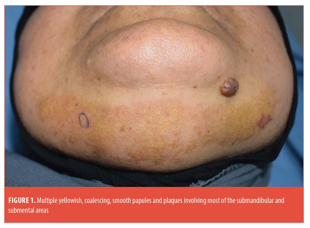
Skin examination showed multiple yellowish, non-tender, smooth, coalescing papules and plaques occupying most of the submandibular and submental areas (Figure 1). General examination revealed generalized edema, puffy eyelids, owed to nephrotic syndrome, and Cushingoid features in the form of moon face, buffalo hump, obese torso and thin, easily bruised skin, all representing the cumulative effect of high-dose steroids taken by the patient. There was no regional lymphadenopathy or organomegaly.
Laboratory findings included elevated levels of serum cholesterol (230 mg/dl) and serum creatinine (1.6 mg/dl). Serum albumin and total protein levels were 2.9 g/dl (normal 3.5 to 5 g/dl) and 5.2 g/dl (normal 6 to 8 g/dl), respectively. Urinalysis disclosed 1+ protein. CBC, ESR, serum triglycerides, blood urea and ANA levels were normal. Abdominal ultrasound showed unremarkable findings.
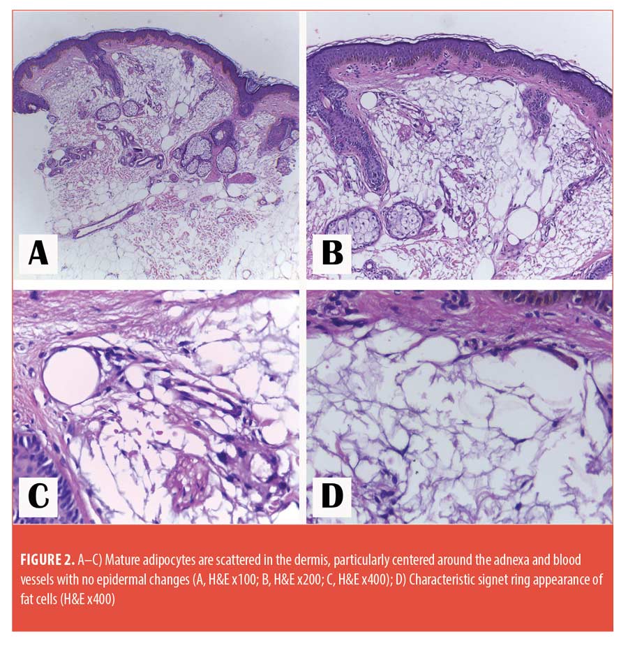
A punch biopsy was taken from one of the yellowish plaques and pathological examination showed lobules of mature adipocytes embedded with collagen fibers in the reticular dermis, with no pathological evidence of xanthoma (Figure 2). Based on these findings, we settled on the diagnosis of NLCS.
NLCS is an uncommon benign hamartomatous cutaneous lesion. The true origin of this rare nevus is obscure and several theories have been proposed to explain its pathogenesis.3 Two clinical types of NLCS are distinguished: the classic (multiple) type and the solitary type. The classic type occurs at birth or during first two decades of life and consists of multiple, soft, pedunculated, skin-colored or yellowish nodules that might coalesce to form plaques. The solitary form usually occurs after the age of 20 years, presents with a single nodular lesion mimicking a skin tag with no particular predilection sites. NLCS is asymptomatic in both types.1
Reported associations include café-au-lait macules, leukodermic spots, overlying hypertrichosis, comedo-like alterations, retractile testis and folliculosebaceous cystic hamartoma.1,3 None of these associations were evident in our patient. However, the clinical appearance is unique and was only reported once before.2 Additionally, the onset of classic NLCS in the elderly along with extension of the lesion across the midline are other rare features observed in our patient.1, 4, 5
NLCS is pathologically characterized by mature adipocytes, irregularly scattered throughout the dermis, particularly centered around the adnexa and blood vessels. Because of this placement, the junction between the dermis and the subcutis is often blurred.
Treatment of NLCS is not indicated except for cosmetic reasons.3 Our patient was offered treatment by carbon dioxide laser, but she refused. Written informed consent was obtained from the patient for publication.
We report this rare case of adult-onset NLCS resembling plane xanthoma with the aim of raising awareness of such morphologically mimicking presentation among dermatologists and highlighting the role of histopathology in confirming the diagnosis.
With regard,
Hussein M. M. Hassab-El-Naby, MD, and Mahmoud A. Rageh, MSc
Affiliations. Both authors are with the Department of Dermatology and Faculty of Medicine at Al-Azhar University in Cairo, Egypt.
Funding. No funding was provided for this article.
Disclosures. The authors report no conflicts of interest relevant to the content of this article.
Correspondence. Mahmoud A. Rageh, MSc; Email: dr.mahmoudrageh@gmail.com
References
- Alsalman HH, Alhallaf RA, Alhuzaimi A, et al. Hairy nevus lipomatosus cutaneous superficialis: a rare presentation. JAAD Case Reports. 2020;6(10):1116–1118.
- Oliveira ALC, Rabay FMO, Elias BLF, et al. Superficial cutaneous lipomatous nevus: report of a case simulating plane xanthoma. Surg Cosmet Dermatol. 2015;7(3 Suppl 1):S53–55.
- Kumaran MS, Narang T, Dogra S, et al. Nevus lipomatosus superficialis unseen or unrecognized: a report of eight cases. J Cutan Med Surg. 2013;17(5):335–339.
- Patil SB, Narchal S, Paricharak M, et al. Nevus lipomatosus cutaneous superficialis: a rare case report. Iran J Med Sci. 2014;39(3):304–307.
- Pasagadugula KV, Prasad PG, Laxmi SJ, et al. Nevus lipomatosus cutaneous superficialis: poses a wholistic health problem: a case report. IOSR Journal. 2015;14(1):14–16.
Social Media Use and Online Reviews for Dermatologists in the United States
Dear Editor:
We assessed the relationship between social media (SM) use and online reviews across seven leading physician review websites (PRW) for dermatologists in the United States (US). PRWs are a popular mode of patient feedback and play a role in guiding patients to initiate care with specialists.1,2 Of note, dermatologists were found to be the most frequently searched of all specialists and generalists in google searches,3 underscoring the role of online reviews in dermatology self-referral and their potential impact in a patient-centered healthcare model. We examined whether dermatologists’ SM use correlates with number of online reviews or average ratings.
A total of 500 dermatologists (10 per US state) were randomly selected using the National Provider Identifier (NPI) registry. Dermatologists’ number of reviews and scores were collected from seven PRWs: Healthgrades.com, Vitals.com, Ratemd.com, Webmd.com, Zocdoc.com, Yelp.com, and Google.com. Dermatologists were assigned an SM score (0–3) as a measure of their total SM use. One point was added for use on each of the three most popular SM platforms: Facebook (FB), Twitter (TW), and Instagram (IG). Multivariable Pearson correlation analysis and two-tailed t-tests were conducted between variables.
Of the cohort 161 (32.2%) had at least one SM account of the three reviewed, the most popular was FB (90.6%) then TW (36.6%) and IG (32.9%). The total number of aggregated online reviews across the seven platforms by SM score was calculated (Table 1). Dermatologists with any SM use had higher number of reviews on average compared to dermatologists with no SM use (192.0 vs. 44.9; P= 1.22×10⁻⁹) but not overall scores. Table 1 shows when subdivided by degree of SM use, increasing SM score corresponded with higher average number of reviews per dermatologist. Of note, SM usage varied significantly by state; the mean SM score was higher for the five most populous versus least populous states (Table 2). The states with the highest average dermatologist SM score were California (2.2.), New York (1.5), Virginia (1.3), Florida (1.2), and Nevada (1.1).
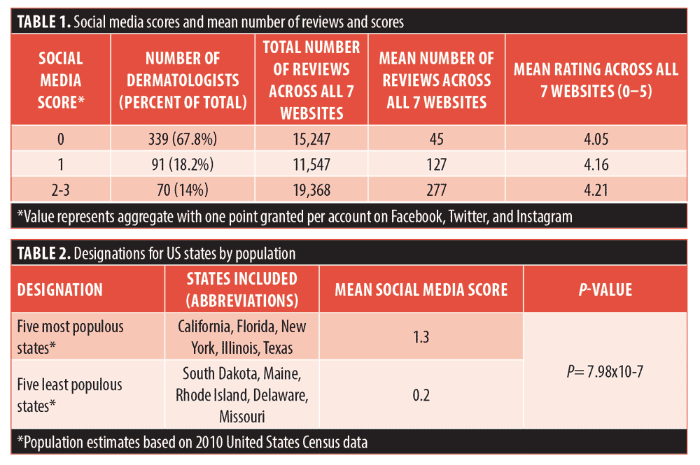
Our results show dermatologists with higher SM use have significantly greater aggregate number of reviews across seven PRWs, suggesting they may be more successful at engaging clientele in ratings and reviews. Moreover, there was a statistically significant relationship between SM score and aggregate number of reviews (P=1.22×10⁻⁹) but notably not between SM score and PRW ratings (Table 1). In concordance with previous studies, we found that mean ratings across all PRWs were high (>4 stars) and there were no significant differences in ratings between websites.4 For improvement in ratings dermatologists may need to focus on the quality of the patient experience rather than invest further resources into SM. While this study focuses on numeric data and whether SM use is associated with differences in ratings and number of reviews, the text of reviews may be a rich data source on what factors impact ratings. Future studies may use that data to identify key common themes and potential strategies for dermatologists to improve the actual rating score.
With regard,
Afsheen Sharifzadeh, MD, and Gideon P. Smith, MD, PhD, MPH
Affiliations. Dr. Sharifzadeh is with the Department of Dermatology at Massachusetts General Hospital in Boston, Massachusetts and the University of Massachusetts Medical School in Worcester, Massachusetts. Dr. Smith is with the Department of Dermatology at Massachusetts General Hospital in Boston, Massachusetts and Harvard Medical School in Boston, Massachusetts
Funding. No funding was provided for this article.
Disclosures. The authors report no conflicts of interest relevant to the content of this article.
Correspondence. Afsheen Sharifzadeh, MD; Email: afsheen.sharifzadeh@gmail.com
References
- Li S, Feng B, Chen M, et al. Physician review websites: effects of the proportion and position of negative reviews on readers’ willingness to choose the doctor. J Health Commun. 2015;20(4):453–461.
- Hong YA, Liang C, Radcliff TA, et al. What Do Patients Say About Doctors Online? A Systematic Review of Studies on Patient Online Reviews [published correction appears in J Med Internet Res. 2019 Jul 18;21(7):e14823]. J Med Internet Res. 2019;21(4):e12521. Published 2019 Apr 8.
- Ransohoff JD, Sarin KY. Referred by Google: mining Google Trends data to identify patterns in and correlates to searches for dermatological concerns and providers. Br J Dermatol. 2018;178(3):794–795.
- Riemer C, Doctor M, Dellavalle RP. Analysis of Online Ratings of Dermatologists. JAMA Dermatol. 2016;152(2):218–219.
Treatment of Periorbital Facial Wrinkles in Female Subjects: A Randomized, Multi-Center, Double-Blinded, Placebo-Controlled, Split-Face Study Evaluating Procedure Pairing of a Peptide Anti-Aging Serum with OnabotulinumtoxinA
Dear Editor:
There is a growing demand for treatments for mimetic wrinkles within dermatology and plastic surgery clinics beyond neuromodulator injections.1 In 2002, the United States (US) Food and Drug Administration (FDA) approved onabotulinumtoxinA (Botox®; Allergan, Dublin, Ireland) as an effective temporary treatment in reducing the appearance of moderate-to-severe mimetic wrinkles.1,2 While onabotulinumtoxinA softens mimetic wrinkles, the overall skin appearance, texture, firmness, and roughness are not directly targeted. Thus, there is a critical need for safe postprocedure pairing cosmeceuticals that can work synergistically with neuromodulators to improve the appearance of facial expression lines as well as enhance overall skin health. A skincare regimen combining in-office procedures with at-home care would enhance patient offerings.
A mimetic line targeted peptide-based treatment serum (LTPS) combining five neuromodulating peptides and gamma aminobutyric acid (GABA) clinically improved the appearance of expression lines in women with mild-to-moderate fine lines and wrinkles after twice-daily application for twelve weeks in a stand-alone study.3 It was hypothesized that this LTPS would provide synergistic effects in pairing with neuromodulators, as well as overall skin health with statistical significance when compared to a placebo serum (PS) paired with a neuromodulator in women with moderate-to-severe crow’s feet wrinkles and fine lines.
In a multicenter, randomized, split-faced, double-blinded, placebo-controlled study with two board certified dermatologists and an aesthetic physician, 36 female subjects between the ages of 35 to 60 with Fitzpatrick Skin Types I to VI, moderate-to-severe crow’s feet wrinkles, and fine lines, who were not pregnant nor planning to become pregnant during the duration of the study nor had been diagnosed with an autoimmune disease, were enrolled. Blinded subjects were randomized to receive the LTPS and PS using a randomization table prepared prior to the start of the study by a non-participating staff member. Subjects were assigned a subject number in numerical order as enrolled and were provided a set of products labeled “R” right and “L” left (depending on the randomization of that subject). Subjects received 10 to 12 units of onabotulinumtoxinA injection (following FDA-approved criteria) in the crow’s feet area bilaterally to have the subject act as their own control. The dosing the subjects received needed to be at a level that would be considered full treatment to demonstrate efficacy thereby raising the difficulty of demonstrating a statistically significant effect. Subjects applied products immediately following neuromodulator injection and twice daily for 12 weeks to the right and left periorbital and lateral canthal region, along with a provided bland moisturizer and sunscreen. Blinded clinical grading was performed using the Modified Griffith’s 10-point scale.4
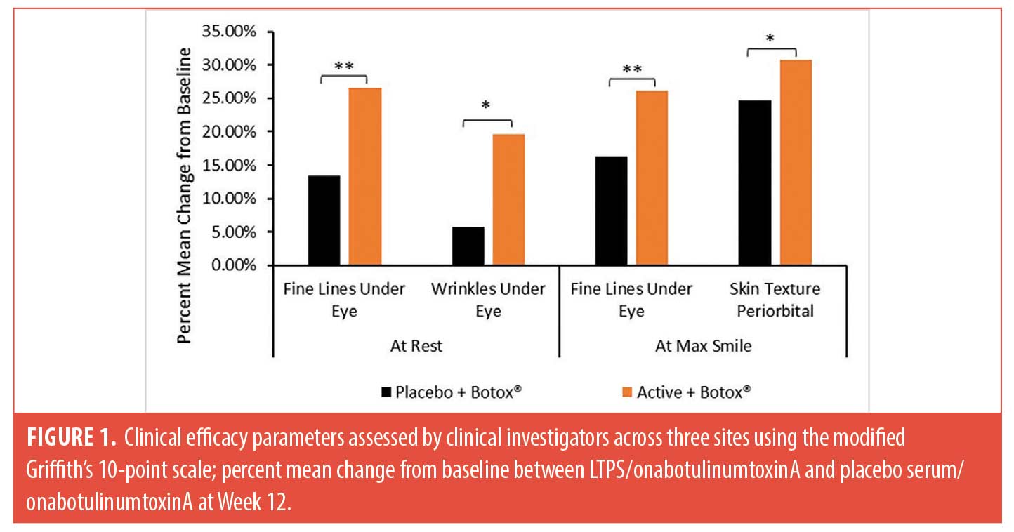
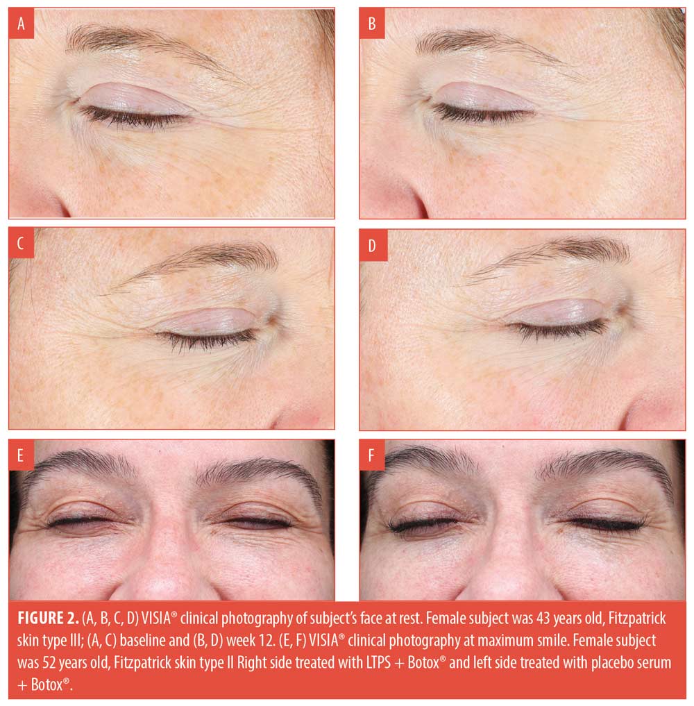
Twenty-nine female subjects with an average age of 49.7 (range 37 to 59 years), across three study centers completed the study. Female subjects were predominantly White (90%), followed by Hispanic (10%), and had primarily Fitzpatrick Skin Type III (59%), followed by Fitzpatrick Skin Type II (21%), Type I (14%), and Type IV (7%). Two sites injected 12 units of onabotulinumtoxinA and one site injected 10 units of onabotulinumtoxinA on both canthal sides across their patients at baseline. LTPS demonstrated superior efficacy by statistically significantly outperforming the PS in improving under-eye fine lines and wrinkles at rest, 26.63 percent (**p<0.01) and 19.60 percent (*p<0.05) respectively, at Week 12 compared to baseline. Similarly, LPTS showed superiority to PS by statistically significantly improving under eye fine lines, 26.12 per (**p<0.01), and periorbital skin texture, 30.82% (*p<0.05), at max smile at Week 12 compared to baseline (Figure 1). Improvement in clinical efficacy parameters, including undereye fine lines and wrinkles and periorbital skin texture, were further demonstrated by VISIA® in standard light; under-eye fine lines and wrinkles at rest (Figure 2A, B, C, D) and at max smile (Figure 2E, F). 3D PRIMOSCR imaging was not performed in this study, however in the stand-alone study PRIMOSCR results indicated an improvement in wrinkle length and total wrinkle count.3
The results of the novel topical LTPS with five neuromodulating peptides and GABA are attributed to blunting of the concentration of acetylcholine at the epidermal surface, thus mitigating horizontal and vertical expression lines.5 Providing a synergistic skincare solution that combines the benefits of in-office neuromodulator procedures and an at-home skincare regimen to enhance patient offerings, improve facial expression lines, and long-term skin health.
With regard,
Alisar S. Zahr, PhD; Sofia Iglesia, MS; Tatiana Kononov, BS, MBA;
Shino Bay Aguilera, DO;
Justin Harper, MD;
and Kimberly Schulz, MD
Affiliations. Dr. Zahr, Ms. Iglesia and Ms. Kononov are with Revision Skincare® in Irving, Texas. Dr. Aguilera is with Shino Bay Cosmetic Dermatology in Fort Lauderdale, Florida, Dr. Harper is with Juvly Aesthetics in Columbus, Ohio, Dr. Schulz is with Infinity Skin Care in Coralville, Iowa.
Funding. Funding was provided by Revision Skincare®.
Disclosures. Dr. Shino Bay Aguilera, Dr. Justin Harper, and Dr. Kim Schulz served as the clinical graders for this clinical trial. Dr. Zahr, Ms. Iglesia, and Ms. Kononov, are employees of Revision Skincare®.
Correspondence. Alisar S. Zahr, PhD; E-mail: azahr@revisionskincare.com
- References
- Lemperle G, Homes RE, Cohen SR, et al. A Classification of Facial Wrinkles. Plast Recontr Surg. 2001;108(6):1735–1750.
- Satriyasa BK. Botulinum Toxin (Botox A) for Reducing the Appearance of Facial Wrinkles: A Literature Review of Clinical Use and Pharmacological Aspect. Clin Cosmet Investig Dermatol. 2019; 12:223–228.
- Nguyen TQ, Zahr AS, Kononov T, et al. A Randomized, Double-blind, Placebo-controlled Clinical Study Investigating the Efficacy and Tolerability of a Peptide Serum Targeting Expression Lines. J Clin Aesthet Dermatol. 2021 May;14(5):14–21.
- Griffiths CE, Wang TS, Hamilton TA, et al. A photonumeric scale for the assessment of cutaneous photodamage. Arch Dermatol. 1992 Mar;128(3):347–351.
- Zahr AS, Koch J, Nguyen TQ, et al. A Novel Blend of Neuromodulating Peptides and Gamma Aminobutyric Acid to Regulate the Release of Acetylcholine. Poster presented at: The American Academy of Dermatology, VMX; April 23–25, 2021.

