 J Clin Aesthet Dermatol. 2021;14(11):35–37.
by John Howard, DO; Cassandra Johnson, MS; and Stanley Skopit, DO, MSE, FAOCD, FAAD
Dr. Howard is with the Department of Dermatology at Larkin Community Hospital in South Miami, Florida. Ms. Johnson is with the Dr. Kiran C. Patel College of Osteopathic Medicine at Nova Southeastern University in Fort Lauderdale, Florida. Dr. Skopit is with the Dermatology Residency Training Program at Larkin Community Hospital in South Miami, Florida.
FUNDING: No funding was provided for this article.
DISCLOSURES: The authors report no conflicts of interest relevant to the content of this article.
ABSTRACT: Kaposi sarcoma (KS) is an angioproliferative vascular neoplasm characterized by the proliferation of endothelial cells that is typically seen in patients with acquired immunodeficiency syndrome (AIDS). There are four major subtypes of KS: classic, African endemic, iatrogenic, and AIDS-associated KS. In rare circumstances, a patient might present with KS that does not fit into any of the four major subtypes and has no identifiable predisposing factors. This case report and review of the literature is presented to serve as a reminder to practitioners to suspect the unexpected when considering cystic or vascular-type lesions on the head and neck.
Key words: Kaposi sarcoma, human immunodeficiency virus-negative Kaposi sarcoma, head and neck Kaposi sarcoma
Kaposi sarcoma (KS) is an angioproliferative vascular neoplasm characterized by the proliferation of endothelial cells.1 There are four major subtypes of KS: classic, African (endemic), iatrogenic, and acquired immunodeficiency syndrome-associated (epidemic) KS.2 Classic KS is characterized by violaceous papules, which progress to form nodules and plaques typically observable on the extremities and found nearly exclusively in elderly men of Mediterranean descent.1 The African subtype, a more aggressive form of KS, is characterized by infiltrative lesions that can progress to visceral involvement.1 Iatrogenic KS is seen in patients with a history of transplantation or an autoimmune disorder who are taking immunosuppressive drugs.1 AIDS-associated KS is the most common subtype, where plaques and nodules commonly affecting the face, extremities, and mucocutaneous tissue can be found.1
In the non-AIDS population, KS is rare, and the frequency differs based on many epidemiologic factors, such as age and sex, as well as immunocompetence. Generally, non-AIDS KS affects older men, presents on the extremities, and is rarely found on the head or neck.3 Additionally, many patients with non-AIDS KS might be on immunosuppressive therapy, making them 150 to 200 times more likely to show symptomatic lesions than the general population.3
Here, we present a case of a 68-year-old immunocompetent Haitian woman who presented with a three-week history of a nodule on her cheek. This case presented with many unique features, making it difficult to definitively categorize it as one of the KS subtypes. Our patient’s sex and ethnicity are inconsistent with the significant male, African, and Mediterranean preponderance of non-AIDS KS. Additionally, the location of our patient’s lesion is uncommon. This unique presentation serves to add to the literature while enhancing the differential diagnoses of the practitioner for cystic and vascular-appearing lesions on the head and neck.
Case Presentation
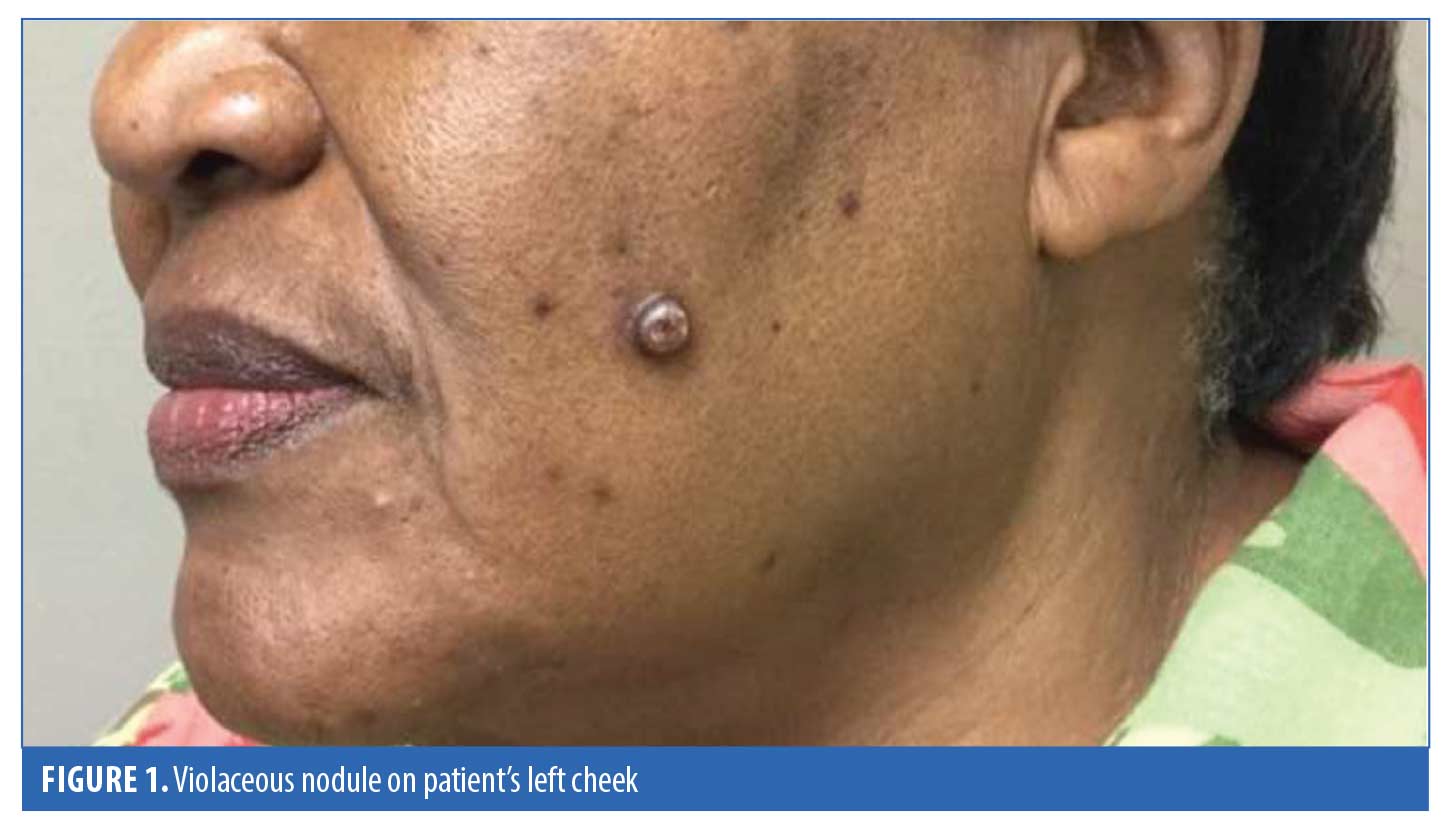
A 68-year-old Haitian woman presented to our dermatology clinic with three-week history of a solitary, 1- by 1-cm violaceous nodule on an erythematous patch located on her left cheek (Figure 1). The patient stated she had developed a “pimple” and had tried to squeeze it, with no improvement. Then, over the subsequent days, the lesion had rapidly grown in size and the patient became concerned, finally seeking medical attention. A physical examination revealed no other suspicious cutaneous or mucosal skin lesions. The patient’s medical history was negative for a prior transplant, immunodeficiency, or autoimmune disease. The travel history for our patient was also negative; she was born in Haiti but came to the United States as a young girl.
A shave biopsy with electrocautery was performed on the lesion and histopathology revealed nodular KS (Figure 2). Immunohistochemical staining for human herpesvirus 8 (HHV8) was strongly positive (Figure 3). Subsequent laboratory findings were negative for human immunodeficiency virus (HIV) 1 and 2, and a complete blood count analysis revealed no abnormalities.
Our patient was then treated with local cryotherapy with liquid nitrogen and experienced complete resolution of the original lesion. During follow-up, six weeks later, postinflammatory hyperpigmentation was noted, and there was no sign of recurrence. 

Discussion
KS has varying clinical and epidemiological differences, allowing for its categorization into separate subtypes. However, HHV8 plays a critical role in the pathogenesis of all KS variants.1 This virus, thought to be an antecedent of KS onset, is universally harbored within the skin lesions.2 The virus has been detected in many bodily fluids, making transmission possible horizontally, sexually, or through organ transplant and blood products.1 In endemic regions, the virus is thought to be acquired by family members in childhood, since the seroprevalence is believed to be as high as 80% with increasing age.1 Although Haiti is not recognized as an endemic region for KS, our patient acquired the virus through some unknown means.
Once infected, HHV8 plays a direct role in the progression to KS through the release of inflammatory and angiogenic factors. The question remains: is KS a reactive process of HHV8-infected cells, or is it a true neoplasm capable of metastasis? Patrikidou et al3 suggested early stages of KS are a reactive polyclonal hyperplasia progressing to a monoclonal process in later stages. It is known that not all HHV8-seropositive individuals develop KS, suggesting viral infection is insufficient in the pathogenesis. Immunocompromised patients are unable to properly fight the HHV8 viral infection, likely leading to progression to KS. However, immunocompetent individuals, such as our patient, can still develop KS. It is possible that a genetic alteration in certain ethnic groups allows for the HHV8 infection to advance. However, not enough research has been done to elicit a clear answer.
The location of the KS lesions is variable depending on the subtype. AIDS-associated KS commonly involves the head and neck, while involvement of this location is rare in patients with the other KS variants.2 A review of literature by Patrikidou et al3 identified 251 cases of non-AIDS KS of the head and neck. In this review, 7.9% of cases were the iatrogenic subtype, with the remainder being either classical or endemic KS. Seleit et al4 described a case of a 42-year-old man with a facial nodule on his right cheek. This patient had no history of immunosuppression and tested negative for HIV. Another case of a 74-year-old man presenting with a violaceous nodular lesion on his left nasal wing also tested negative for HIV.5 These cases are similar to our case, since both had no history of immunosuppression and presented with only solitary lesions of KS.
KS has classically been a male-dominated disease, with each subtype having a different prevalence of male to female cases ratio. Classical KS, the most male-dominated variant, occurs at a male to female ratio of 17:1.1 The review of non-AIDS KS of the head and neck by Patrikidou et al3 reported a male to female ratio of 3.1:1, differing greatly from the classic KS ratio. Mohanna et al6 presented a case of a 71-year-old Quechua woman with a painless papules on her hard palate found to be KS on biopsy. The patient had no history of immunosuppression and was negative for HIV. Bottler et al7 described a case of a 76-year-old Caucasian woman with a nodular growth on the dorsal aspect of her tongue. Her medical history was also insignificant and serologic testing was negative for HIV. Both cases reported oral lesions in women with no history of immunosuppression. Notably, both women were not of sub-Saharan African descent, as was our patient. Clinical diagnosis and classification of a KS into one of its subtypes is a diagnostic challenge when lesions present in uncharacteristic locations in women with no risk factors and of ethnicities not associated with KS.
A thorough review of the literature was completed to identify cases of KS that did not fit the epidemiological characteristics of classical KS or the other subtypes. Only 14 cases were identified and the characteristics for all the cases are summarized in Table 1. We only included cases of immunocompetent patients without Mediterranean descent (including the countries of Cyprus, Greece, Lebanon, Syria, Egypt, Italy, Spain, Palestine, Israel, Turkey, and Jordan), just as in our case. Many of the cases included were reported to be classical KS according to the author(s); however, we included such cases in the table because they had unique characteristics and were not representative of the typical epidemiology of classical KS. While more subtypes of KS might exist, it is possible some cases of classic KS do not fit any of the typical epidemiologic characteristics.
The diagnosis of KS can be made through hematoxylin and eosin staining alone. With nodular KS, the specimen might demonstrate a nodule composed of fascicles of parallel spindle cells, erythrocytes between spindle cells, eosinophilic globules, mitoses, and hemosiderin (Figure 2). The irregular vascular spaces between fascicles are commonly referred to as “slit-like” spaces. In our case, the HHV8 immunohistochemical staining added to the diagnosis of KS by demonstrating antibodies to viral particles in the nucleus (Figure 3). Although immunohistochemical staining is not necessary for the diagnosis of KS, it can be useful in differentiating KS from Kaposiform hemangioendothelioma, moderately differentiated angiosarcoma, and spindle cell hemangioma.
First-line therapies for limited cutaneous KS include cryotherapy, intralesional vinblastine, radiotherapy, and alitretinoin gel.8 In the case of our patient, cryotherapy was used and led to a cosmetically appealing outcome. She was treated with a double freeze–thaw cycle of eight seconds per cycle. For more resistant limited cases, extensive cutaneous involvement, systemic KS, or tumor-associated lymphedema, the clinician should consider more aggressive treatment modalities, such as liposomal anthracyclines, taxanes, and interferon-alpha therapy.8 Other therapies on the horizon for the treatment of KS include antiangiogenic agents, antiviral agents, orally active matrix metalloproteinase inhibitors, tyrosine kinase inhibitors, and interleukin-12 inhibitors.8
References
- Fatahzadeh M. Kaposi sarcoma: review and medical management update. Oral Med Oral Pathol Oral Radiol. 2012;113(1):2–16.
- Ramírez-Amador V, Anaya-Saavedra G, Martinez-Mata G. Kaposi’s sarcoma of the head and neck: a review. Oral Oncol. 2010;46(3):135–145.
- Patrikidou A, Vahtsevanos K, Charalambidou M, et al. Non-AIDS Kaposi’s sarcoma in the head and neck area. Head Neck. 2018;31(2):260–268.
- Seleit I, Attia A, Maraee A, et al. Isolated Kaposi Sarcoma in two HIV negative patients. J Dermatol Case Rep. 2011;5(2):24–26.
- Masala MV, Montesu MA, Cottoni F. A rare observation: Kaposi’s sarcoma in HIV-negative spouses. J Eur Acad Dermatol Venereol. 2018;20(10):1355–1356.
- Mohanna S, Francisco B, Ferrufino JC, Sanchez J, Gotuzzo E. Classic Kaposi’s sarcoma presenting in the oral cavity of two HIV-negative Quechua patients. Med Oral Patol Oral Cir Bucal. 2018;12(5):365–368.
- Bottler T, Kuttenberger J, Hardt N, Oehen H-P, Baltensperger M. Non-HIV-associated Kaposi’s sarcoma of the tongue: case report and review of the literature. Int J Oral Maxillofac Surg. 2007;36(12):1218–1220.
- Lebwohl MG, Heymann WR, Berth-Jones J, Coulson I. Treatment of Skin Disease: Comprehensive Therapeutic Strategies. 5 ed. Amsterdam, the Netherlands: Elsevier; 2017.

